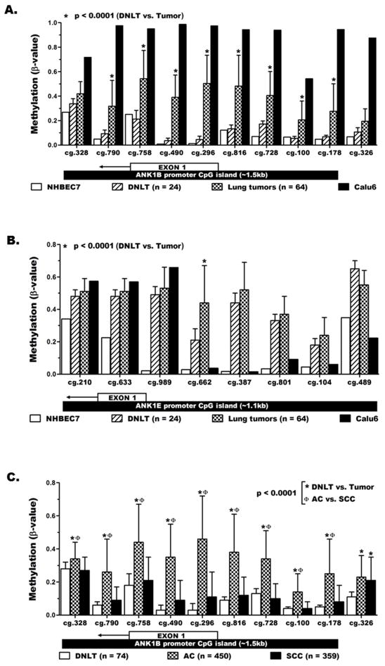
Figure 4. Quantitative validation of ANK1 methylation in lung cancer.
The methylation level (mean ± SD) of (A) ANK1B and (B) ANK1E promoter CpG islands was quantitatively determined using whole genome methylation data from the HumanMethylation450 beadchip (HM450K). The location of each probe with respect to the promoter CpG island region and first exon of the specific transcript variant is depicted below the x-axis labels. C) HM450K data for large lung tumor and normal samples from the publicly available TCGA database validated the tumor-specific (not present in normal lung) methylation of ANK1B promoter in lung cancer and revealed its strong association with lung adenocarcinoma (AC) than squamous cell carcinoma SCC.

