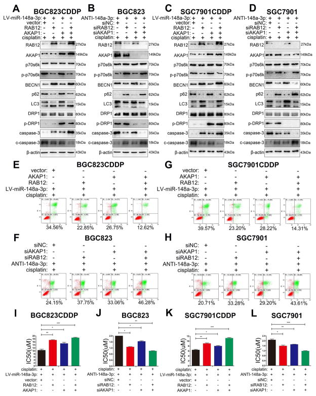
Figure 8. miR-148a-3p enhanced CDDP cytotoxicity by simultaneously inhibiting AKAP1 and RAB12 expression in GC cells.
miR-148a-3p reconstituted CDDP resistant cells (BGC823CDDP, SGC7901CDDP) with RAB12, AKAP1 single or combined overexpression or miR-148a-3p inhibited CDDP sensitive cells (BGC823, SGC7901) with RAB12, AKAP1 single or combined knockdown were tested with 48h CDDP treatment. (A–D) Western blot analysis of RAB12, AKAP1, p70s6k, p-p70s6k(Thr389), BECN1, p62, LC3, DRP1, p-DRP1(Ser637), caspase-3, and cleaved caspase-3 protein levels. (E–H) Flow cytometry analysis of cell apoptosis. (I–L) CDDP IC50s calculated using CCK-8. CDDP concentrations: same as in Figure 2. β-actin: internal control. Graph represents mean ± SEM; *P<0.05, **P<0.01, ***P<0.001.

