Fig. 6.
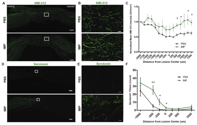
IMPs treated animals have higher axonal density both within and caudal to the lesion site. (A) Representative images of 30 μm thick mid-sagittal sections of PBS or IMPs treated animals 26 weeks post SCI stained with SMI-312. Scale bar = 250 μm (B) Enlarged images from the outlined lesion areas. Scale bar = 20 μm. (C) SMI-312 staining intensity is increased caudal to the lesion site in IMP-treated mice. (n = 4 mice for PBS and n = 3 mice for IMPs group. *p < 0.05 by unpaired Student’s t-test) (D) Representative images of 16 μm thick mid-sagittal sections of PBS or IMPs-treated animals 13 weeks post SCI stained for serotonin (5-HT). Scale bar = 250 μm (E) Enlarged images from the outlined lesion areas. Scale bar = 10 μm. (F) 5-HT+ fibers are more numerous within and immediately rostral to the lesion core in IMP-treated animals as compared to control. (n = 8 mice per group) All data are presented as mean ± SEM. *p < 0.05, **p < 0.01 groups compared by Student’s t-test. Distances rostral to the lesion center are defined as negative numbers, while distances caudal to the lesion center are defined as positive.

