Fig. 1.
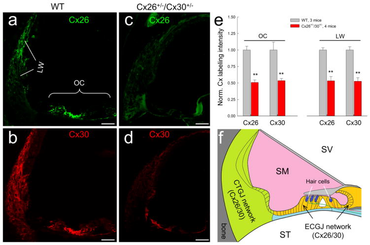
Gap junction network in the cochlea and Cx26 and Cx30 expression in the Cx26+/−/Cx30+/− mouse cochlea. a–d: Immunofluorescent staining for Cx26 (green) and Cx30 (red) in the cochlea in Cx26+/−/Cx30+/− and WT mice. Both Cx26 and Cx30 labeling is visible in the Cx26+/−/Cx30+/− mouse cochlea but their intensities are weaker in comparison with those in WT mice. Scale bar: 50 μm. e: Quantitative measure of Cx26 and Cx30 labeling in the organ of Corti (OC) and the cochlear lateral wall (LW) in WT mice and Cx26+/−/Cx30−/+ mice. The intensity of labeling was normalized to the average value in WT mice. The expression of Cx26 and Cx30 in Cx26+/−/Cx30+/− mice was significantly reduced. **: P > 0.001, t-test. f: Schematic drawing of two gap junctional (GJ) networks in the cochlea: epithelial cell GJ (ECGJ) network in the cochlear sensory epithelium and connective tissue GJ (CTGJ) network in the cochlear lateral wall. Hair cells have neither gap junction nor connexin expression. SV, scala vestibuli; SM, scala media; ST, scala tympani.

