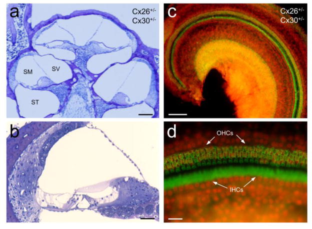
Fig. 4.
Normal development of the cochlea and no hair cell loss in Cx26+/−/Cx30+/− mice. a–b: Cross-sections of the Cx26+/−/Cx30+/− mouse cochlea. The cochlea demonstrates normal shape and has no apparent cell degeneration. SV, scala vestibuli; SM, scala media; ST, scala tympani. c–d: whole-mounting of the cochlear sensory epithelium in Cx26+/−/Cx30+/− mice. Inner hair cells (IHCs) and outer hair cells (OHCs) were stained with phalloidin-Alexa 488 (green) and the cell nuclei were labeled with propidium iodide (PI, red). Mice were 2–3 months old. Scale bar: 100 μm in a & c, 40 μm in b, and 20 μm in d.

