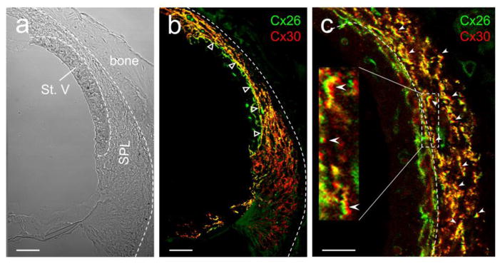
Fig. 8.
Co-expression of Cx26 and Cx30 and EP generation in the cochlear lateral wall. The cross-section of the mouse cochlear lateral wall was stained for Cx26 (green) and Cx30 (red) in immunofluorescent staining. Empty triangles in panel b indicate heterozygous coupling between stria vascularis (basal cell layer) and spiral ligament (fibrocyte I cells). White errors in panel c indicate co-labeling of Cx26 and Cx30 at the same GJ plaques between fibrocyte cells in the spiral ligament. Most of GJ plaque-punctates have Cx26 and Cx30 co-labeling and show yellow color in the over-lap image. St. V, stria vascularis; SPL, spiral ligament. Scale bar: 20 μm in a & b and 10 μm in c.

