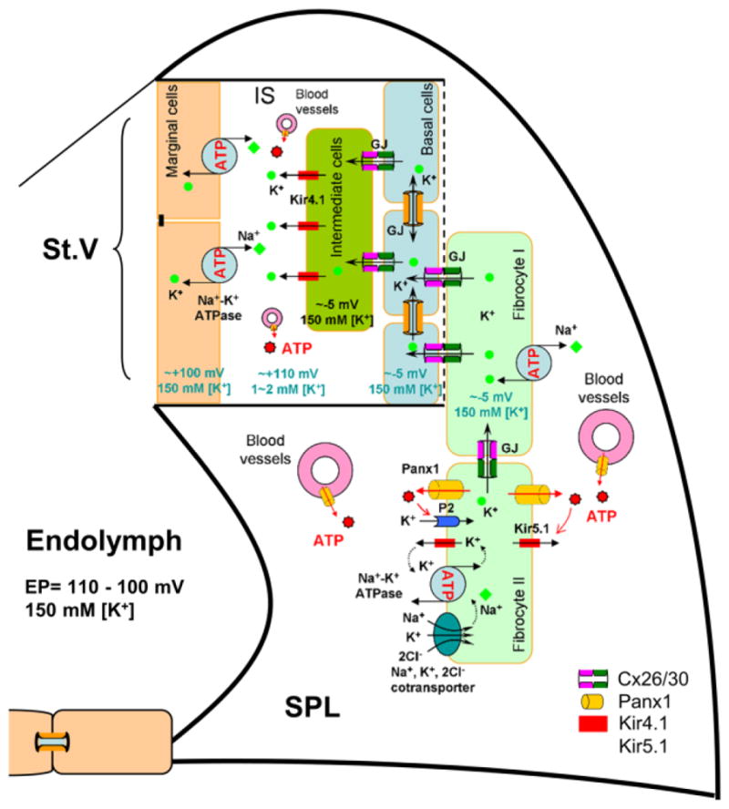
Fig. 9.

Schematic drawing of EP generation in the cochlear lateral wall. Based on the “two-cell” model, EP is generated by Kir4.1 in the apical membrane of intermediate cells in conjunction with Kir5.1 channels, Na+/K+ ATPases, and Na+, K+, 2Cl−-cotransporters in the fibrocytes through GJ coupling [40,45]. Heterozygous couplings of gap junctions between fibrocute I and II cells in the spiral ligament (SPL), between fibrocyte I cells and base cells in the stria vascularis (St. V), and between base cells and intermediate cells are important for eventually positive EP generation. MC: marginal cell; BC: basal cell; FC: fibrocyte; IC: intermediate cell; IS: intrastrial space; GJ: gap junction.
