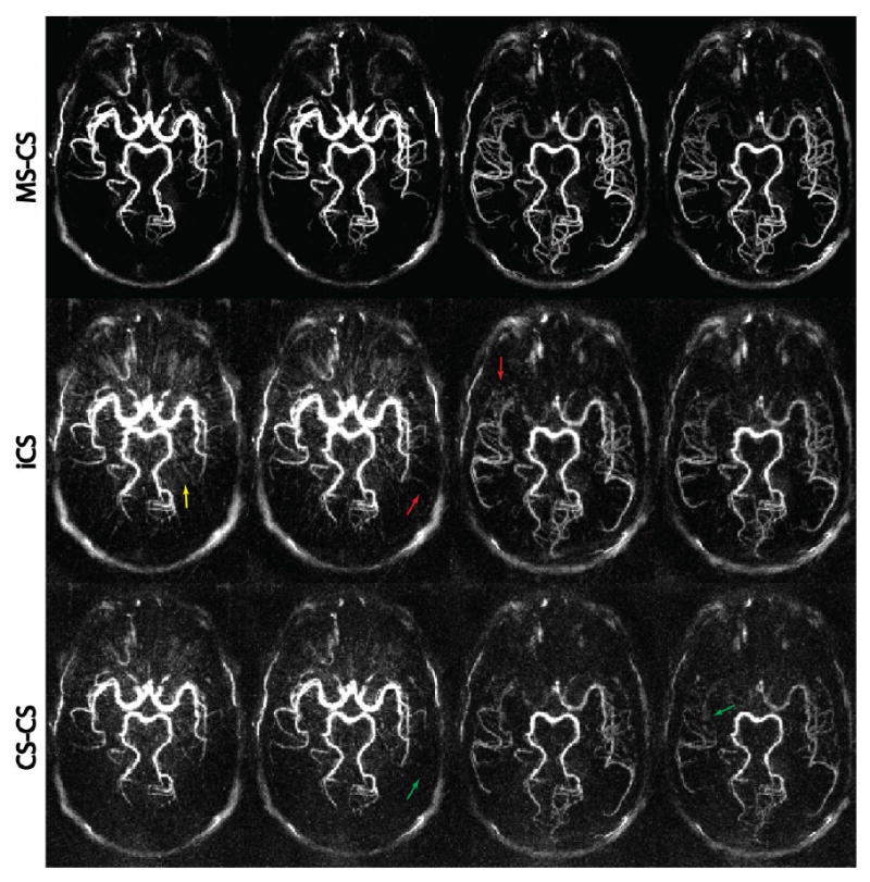
Figure 4.

Comparison of the three CS reconstruction strategies. From top to bottom, each row represents axial MIP images at four time frames reconstructed with MS-CS, iCS, and CS-CS, respectively. Severe streaking artifacts and high noise level are clearly visible on the iCS and CS-CS reconstructions, while the proposed MS-CS reconstruction provides cleaner and sharper images. All images were normalized by its maximum intensity and displayed at the same window level.
