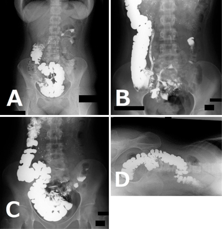
Figure 3.
Gastrointestinal transit X-ray study with oral barium. (A) The upright image obtained 3 hours after the oral administration of barium shows the cecum displaced medially with 180 degrees of torsion. (B) The supine image shows the cecum and ascending colon located in a normal anatomical position. (C) The image shows the cecum and ascending colon displaced toward the pelvic cavity when the patient changed from the supine position to the upright position on the fluoroscopic table. (D) A left lateral decubitus image showing that the entire right sided colon dropped medially beyond the vertebral line.

