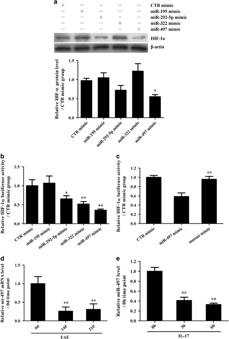
Figure 4.
Effects of potential miRNAs on hypoxia inducible factor-1α (HIF-1α) in interleukin (IL)-17-stimulated astrocytes. (a) Primary mouse astrocytes were transfected with different mimics of miR-195, miR-292-5p, miR-322, miR-497 and CTR, and the protein level of HIF-1α in the astrocytes was detected with western blotting 48 h after transfection. *P<0.05 versus the CTR mimic group. (b) HEK293T cells were transfected with a mixture of pGL3-Promoter/HIF-1α and pRL-SV40 as well as one of the miRNA mimics (miR-195, miR-292-5p, miR-322, miR-497 or CTR). Subsequently, the luciferase activity of the HIF-1α 3′UTR reporter was measured 48 h after transfection. *P<0.05; **P<0.01 versus the CTR mimic group. (c) HEK293T cells were transfected with a mixture of pGL3-Promoter/HIF-1α and pRL-SV40 as well as a miR-497 mimic or miR-497 mutant mimic. Then, the luciferase activity of the HIF-1α 3′UTR reporter was examined 48 h after transfection. **P<0.01 versus the miR-497 mimic group. (d, e) Real-time PCR was used to measure the level of miR-497 in the brain tissues of mice on 0 d, 14 d and 21 d after experimental autoimmune encephalomyelitis (EAE) induction (d, **P<0.01 versus the 0 d time point) or in the astrocytes at 0, 3 and 6 h after IL-17 stimulation (e, **P<0.01 versus the 0 h time point). The results from one representative experiment out of three are shown. Data are shown as the means±s.d. (n=3 in each group).

