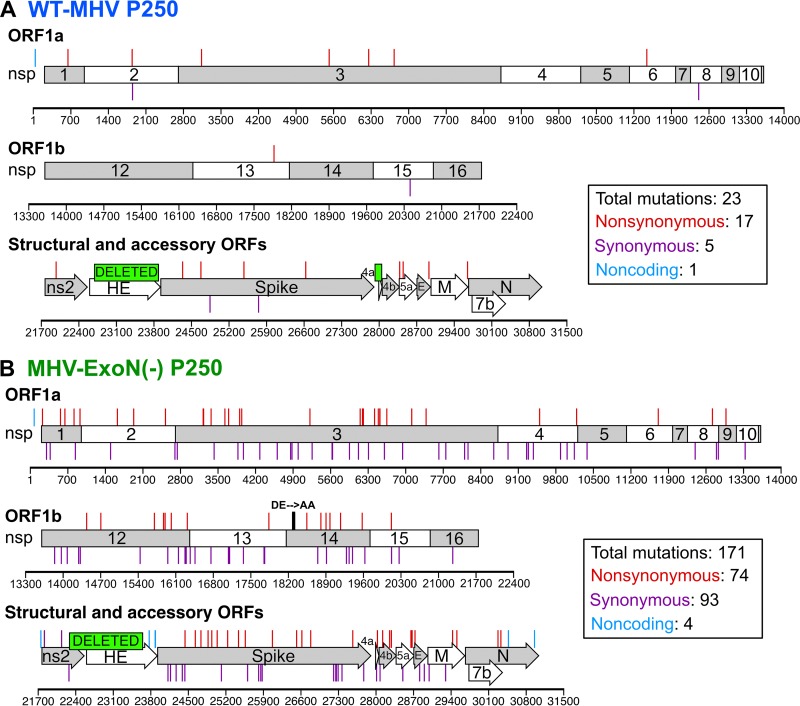
FIG 3 .
Mutations within P250 viruses. The mutations shown were present at >50% by di-deoxy sequencing at passage 250 in WT-MHV (A) and MHV-ExoN(-) (B). Nonsynonymous mutations (red), noncoding mutations (cyan), and deletions (green boxes) are plotted above the schematic, and synonymous mutations (purple) are plotted below the schematic.

