
FIG 5 .
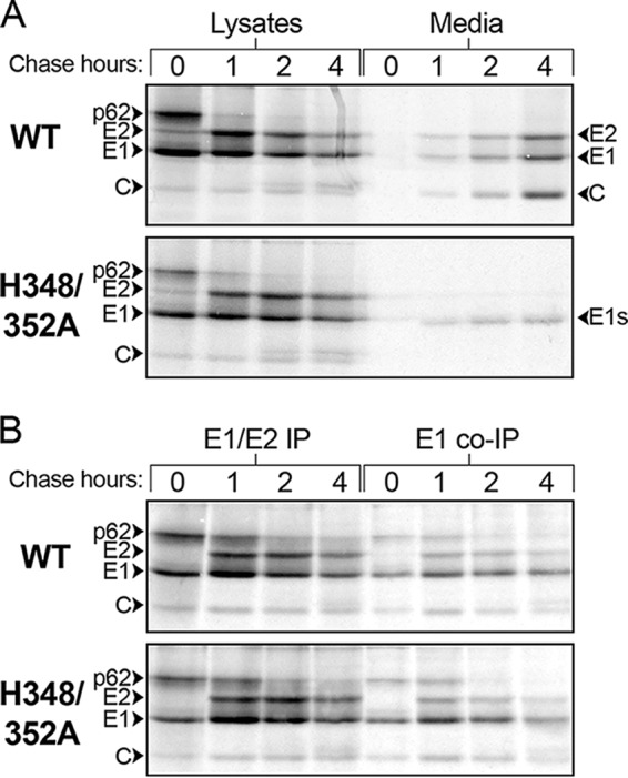
Assembly and dimer stability of WT and mutant viruses. (A) Virus assembly. BHK cells were infected with WT SFV or the H348/352A mutant for 5 h, pulse-labeled for 30 min with [35S]methionine/cysteine, and chased for the indicated times. At each time point, the cells were lysed and immunoprecipitated with a pAb (to E1 and E2), and the virus in the chase medium was retrieved by IP in the absence of detergent. Samples were analyzed by SDS-PAGE and fluorography. The positions of the viral structural proteins and E1s, the soluble truncated form of E1, are indicated. (B) Dimer stability. BHK cells were infected, pulse-labeled, and chased as described for panel A. The cell lysates were immunoprecipitated with a pAb to E1 and E2 or a MAb to E1. The results shown in panels A and B are representative of 2 independent experiments.