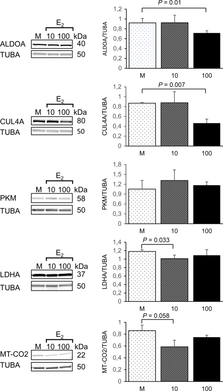
Figure 5.

Validation of the proteomic results by semi‐quantitative Western blotting in primary human muscle cells. Representative images of the Western blots from cell culture experiments with human muscle primary cells. Quantification of the immunoblots was performed with three independent cell experiments exposed to the statistical testing using independent samples t‐test. Data are presented as mean ± standard deviation. M = mock, 10 = 10 nm E2, 100 = 100 nm E2.
