
Figure 2.
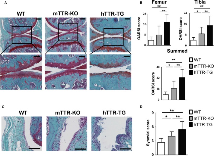
Cartilage and synovial lesions in surgically induced osteoarthritis (OA) in wild‐type (WT), mTTR‐KO, and hTTR‐TG mice. (A) Histological features of knee cartilage 10 weeks after OA surgery (safranin O/fast green; upper: 10×, scale bars = 200 μm, lower: 20×, scale bars = 100 μm). OA was surgically induced by destabilizing the medial meniscus in the right knee joints of 4‐month‐old male mice. (B) Histological OA grade was based on Osteoarthritis Research Society International (OARSI) scoring system. Cartilage scores were significantly higher in hTTR‐TG mice (n = 26) 10 weeks after surgery than in WT (n = 20) and mTTR‐KO mice (n = 22). (C) Histological changes in synovium 10 weeks after OA surgery (scale bars = 100 μm). (D) The synovial score was obtained with Krenn's synovitis scoring system. Synovial scores were significantly higher in hTTR‐TG mice (n = 26) 10 weeks after surgery than in WT (n = 20) and mTTR‐KO mice (n = 22). **P < 0.01 and *P < 0.05.