Figure 1.

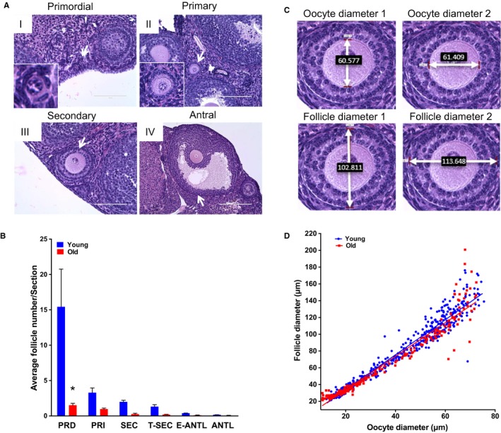
Reproductive aging in CB6F1 mice is associated with reduced ovarian reserve and altered follicle growth dynamics. (A) Representative hematoxylin and eosin (H&E)‐stained ovarian tissue sections demonstrate follicle‐stage classes according to histologic morphology (see Fig. S1 for additional images). White arrows highlight the particular follicle class. Insets show magnified images of primordial and primary follicles. Scale bars are (I–III) 100 μm and (IV) 200 μm. (B) Graph showing the average number of each follicle class per ovarian section (every fifth section of serially sectioned ovaries was counted; N = 3 ovaries from reproductively young and old mice). PRD: primordial; PRI: primary; SEC: secondary; T‐SEC: transitioning secondary; E‐ANTL: early antral; ANTL: antral. Two‐way anova was performed; asterisks denote P < 0.001. (C) Images showing how oocyte and follicle diameters were determined by the mean of two perpendicular measurements on H&E‐stained ovarian tissue sections at 40× magnification. (D) Mean oocyte and follicle diameters were plotted for each individual growing follicle (primary–transitioning secondary). Each dot shows the mean oocyte diameter on the x‐axis, and its corresponding follicle diameter on the y‐axis. For each ovary, a minimum of 50 follicles in each stage were measured. Pearson's correlation and linear regression were performed. R 2 for young and old were 0.9455 and 0.9256, respectively. The slopes between age cohorts were significantly different (P = 0.039).
