Figure 1.

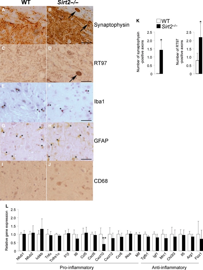
Sirt2−/− mice present axonal degeneration but no neuroinflammation at 13 months of age. Longitudinal and transversal sections of the dorsal spinal cord in WT (A, C, E, G, I) and Sirt2−/− mice (B, D, F, H, J) at 13 months of age (n = 5/genotype) processed for synaptophysin (A–B), RT97 (C–D), Iba‐1 (E–F), GFAP (G‐H), and CD68 (I‐J). Arrows indicate synaptophysin (A–B) and RT97 (C–D) accumulation in axonal swellings. Small stars indicate Iba‐1+ cells (E‐F) and GFAP + cells (G‐H). Scale bar = 25 μm. (K) Quantification of synaptophysin and RT97 accumulation in axonal swellings in 1‐cm‐long longitudinal sections of the dorsal spinal cord in WT and Sirt2−/− mice. The number of abnormal specific profiles was counted at every 10 sections for each stain. At least three sections corresponding to the dorsal columns of the spinal cord were analyzed per animal and per stain. (L) Relative mRNA levels of Nfκb1, Nfκb2, Ikbkb, Tnfα, Il1β, Il6, Cxcl10, Cxcl5, Mif, Tnfrs1α, Ccl5, Ccr6, Cxcl12, Tgfβ1, iNos, Igf1, Mrc1, Chi3 l3, Il5, Arg1, and Fizz1 in spinal cord from WT and Sirt2−/− mice at 13 months of age (n = 8/genotype). Data represent mean ± SD. Statistical analysis was carried out with Student's t‐test (*P ≤ 0.05, **P ≤ 0.01).
