. 2017 Aug 3;8(11):7521–7527. doi: 10.1039/c7sc02622f

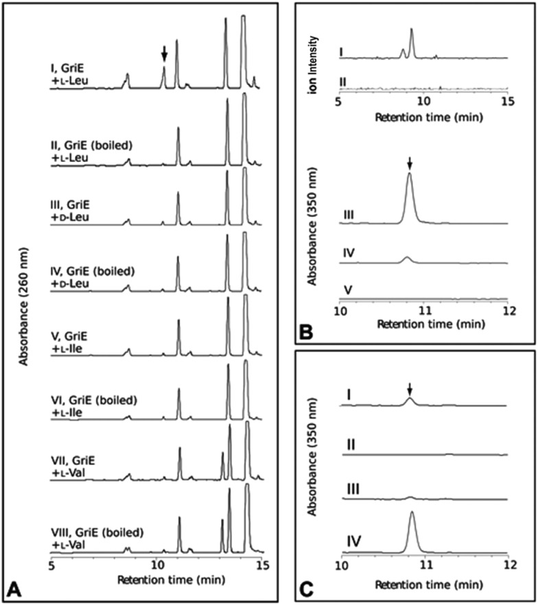
Fig. 3. In vitro analysis of the purified proteins GriE, GriF and GriH. (A) GriE: (I) 5-hydroxyleucine was detected by LC-MS analysis after the incubation of GriE with l-leucine and α-ketoglutarate. (II) Heat inactivated GriE did not yield any 5-hydroxyleucine. 5-Hydroxyleucine was characterized as described in the ESI, Sections 3.10 and 3.11. No reaction products were obtained using d-leucine, l-isoleucine or l-valine as substrates (III–VIII). (B) The extracted ion chromatogram (m/z 128) corresponding to (3R,5S)-3-methyl-Δ1-pyrroline-5-carboxylic acid in positive ion mode (I and II). GriF: (I) the formation of (3R,5S)-3-methyl-Δ1-pyrroline-5-carboxylic acid was observed upon the incubation of GriF with NAD+ and 5-hydroxyleucine. (II) Heat inactivated GriF did not yield any (3R,5S)-3-methyl-Δ1-pyrroline-5-carboxylic acid. (III) The incubation of GriF with ProC from E. coli, 5-hydroxyleucine and NAD+ resulted in the formation of (2S,4R)-4-methyl-proline. (IV) The reaction of GriF in the absence of ProC resulted in trace amounts of (2S,4R)-4-methyl-proline, probably due to a nonenzymatic reduction due to the presence of DTT as a reductant. (V) No formation of (2S,4R)-4-methyl-proline could be detected in the absence of GriF. (C) GriH coupled to GriF: (I) the formation of (2S,4R)-4-MePro was observed upon the incubation of the enzymes with NADH, NAD+, 5-hydroxyleucine and cell extract of S. lividans. (II) Removal of GriF abolished the formation of (2S,4R)-4-MePro. (III) Removal of GriH lead to only trace amounts of (2S,4R)-4-MePro. (IV) The removal of cellular extract from the reaction resulted in higher amounts of (2S,4R)-4-MePro. The stereochemistry of the produced (2S,4R)-4-MePro was confirmed by Advanced Marfey’s Method with d-FDLA derivatized samples.

Fig. 3