. 2017 Aug 3;8(11):7521–7527. doi: 10.1039/c7sc02622f

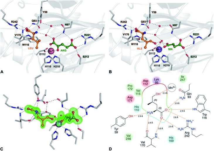
Fig. 5. Crystal structures of the GriE substrate and product complexes. (A) Structure of the enzyme co-crystallized with Co2+, l-leucine (LEU, orange) and α-ketoglutarate (AKG, green). (B) Active site of the Mn2+-containing enzyme with the reaction products (2S,4R)-5-hydroxyleucine (HL5, orange) and succinate (SIN, green). (C) |F O F C| difference electron density of 5-hydroxyleucine and succinate, contoured at 3σ. (D) Ligand interaction diagram for the reaction product (2S,4R)-5-hydroxyleucine. Hydrogen bonds are shown as orange arrows, dashed for side chain-ligand bonds and solid for main chain-ligand bonds. Metal coordination is shown as an orange line. The binding pocket and residues forming it are highlighted according to their physicochemical properties (green: hydrophobic, blue: polar, red: negatively charged, and purple: positively charged).

Fig. 5