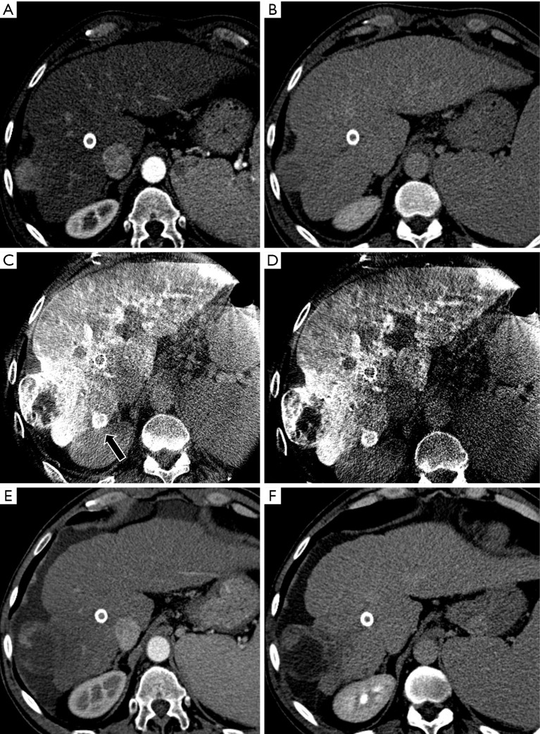
Figure 2.
A case of an intraprocedural CBCT diagnosis of occult nodule in a patient with a S6 (40 mm) exophytic HCC. MDCT arterial (A) and delayed phase (B) shows the presence of the main nodule, with no satellite nodules. Intraprocedural CBCT arterial (C) and venous phases (D) demonstrate a satellite nodule (black arrow) with typical behaviour that was intra-procedurally treated. One-month MDCT demonstrate complete response (E,F). HCC, hepatocellular carcinoma; MDCT, multidetector computer tomography.

