Figure 1.
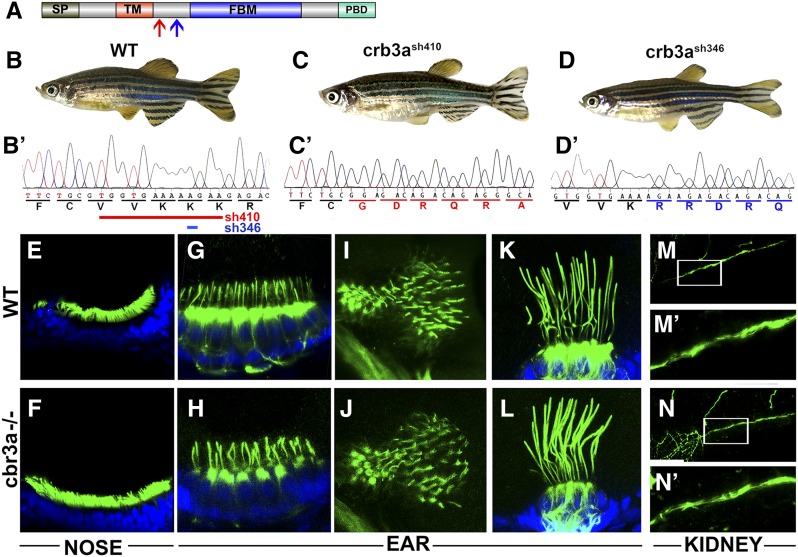
crb3a mutant phenotype. (A) Schematic of Crb3a protein domain structure. Signal peptide (SP); transmembrane domain (TM); FERM-binding motif (FBM); and PDZ-binding domain (PBD) are indicated. Red arrow indicates the start of the frameshift in crb3a−/−sh410 mutant allele; blue arrow, the start of frameshift in crb3a−/−sh346 allele. (B–D) External phenotypes of wild-type (WT) (B), crb3a−/−sh410 homozygous mutant (C), and crb3a−/−sh346 homozygous mutant (D) adult zebrafish. (B’–D’) Sequences of wild-type (B’), and two mutant alleles: crb3a−/−sh410 (C’), and crb3a−/−sh346 (D’). Deletions in crb3a−/−sh410 (red line) and crb3a−/−sh346 (blue line) mutants are indicated in (B’). (E–N’) Images of wild-type and crb3a−/− mutant embryos stained with anti-acetylated tubulin antibody (in green) and counterstained with DAPI (in blue) at 5 days postfertilization: olfactory placode (E and F); anterior macula (G and H); posterior macula (I and J); lateral crista (K and L); and pronephros (M and N). (M’ and N’) are enlarged images of pronephric cilia shown in (M and N).

