Figure 2.

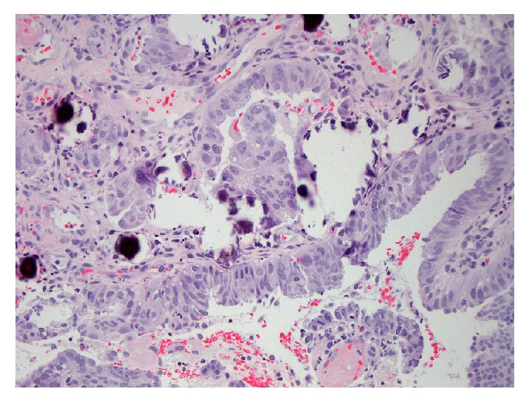
Hematoxylin and eosin staining shows fragments of large atypical cells with focal gland formation consistent with an adenocarcinoma.

Hematoxylin and eosin staining shows fragments of large atypical cells with focal gland formation consistent with an adenocarcinoma.