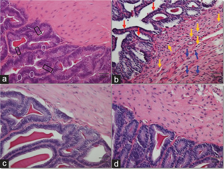
Figure 1.

Histological alterations in the tissue of the seminal vesicles. Black rectangles mark typical pseudostratified tall columnar epithelial cells. White circles surround typical basal cells in the epithelium. Yellow arrows point at the vacuolation observed in the cytoplasm of inner circular muscle cells. Blue arrows indicate typical hyperchromatic nuclei of muscle cells. Red arrows show the shrinking of the epithelial cells. Original magnification: ×400. The scale bar is 30 μm. (a) Control group; (b) DM group, untreated diabetic animals; (c) DM/Eda group, diabetic animals treated daily with edaravone 10 mg kg−1, i.p.; (d) DM/Tau group, diabetic animals treated daily with taurine 500 mg kg−1, i.p.
