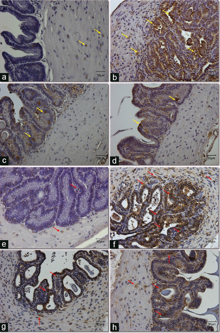
Figure 3.

Expression and localization of apoptotic protein, cleaved caspase-3 (a–d) and lipid peroxidation marker, 4-HNE (e–h) in seminal vesicles section from all groups. (a–d) Yellow arrows point at the positive cells stained with brown color for the cleavedcaspase-3 antibody. (e–h) Red arrows point at the positive cells stained with brown color for the 4-HNE antibody. Original magnification: ×400. The scale bar is 30 μm. (a/e) Control group; (b/f) DM group, untreated diabetic animals; (c/g) DM/Eda group, diabetic animals treated daily with edaravone 10 mg kg−1, i.p.; (d/h) DM/Tau group, diabetic animals treated daily with taurine 500 mg kg−1, i.p. 4-HNE: 4-hydroxy-2-nonenal.
