
Figure 2.
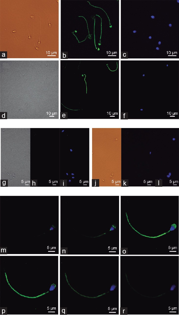
Subcellular localization of the active form of AMPK (phospho-Thr172-AMPK) in human spermatozoa. Phospho-Thr172-AMPK was detected in spermatozoa with the secondary antibody Alexa Fluor 488 goat anti-rabbit immunoglobulin G. Sperm were stained in green with anti-phospho-Thr172-AMPK antibody while sperm nuclei were stained with DAPI (blue). Images from (a–f): Phospho-Thr172-AMPK staining was visualized using epifluorescence (a–c), or confocal microscopy (d–f). Left panels (a and d) show bright field images, central panels (b and e) show IgG-Alexa 488 images and right panels (c and f) represent DAPI images. Images from (g–l) show negative controls: (g–i) Spermatozoa were preincubated with phospho-Thr172-AMPK specific blocking peptide (5X); (j–l) spermatozoa were incubated only with the secondary antibody Alexa Fluor 488 and primary antibody was omitted. Serial images (m–r) of confocal microscopy were obtained from a unique spermatozoon using different focus depths (5 μm thickness) and showing the overlapping IgG-Alexa 488 (green) and DAPI (blue) staining after incubation with the anti-phospho-Thr172-AMPK antibody. Images are representative of n = 6 experiments. Scale bars (5–10 μm) are indicated.