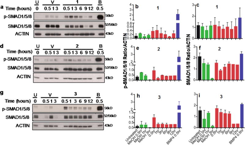
Figure 5. Activation of the canonical BMP signaling pathway.

Immunoblotting analysis of C33A-2D2 cells untreated (U) or treated with: Vehicle (V, 0.034%–0.038% MeOH), 10ng BMP4 (B) or 1 (a), 2 (d), or 3 (g) for 0.5 to 12hrs as indicated. Proteins were separated on 10% or 12% PAGE gels and immunoblotted with antibodies to phosphorylated SMAD1/5/8 (p-SMAD1/5/8) or total SMAD1/5/8. ACTIN was used as loading control. Quantification of protein signal for p-SMAD1/5/8 (b,e,h) and SMAD1/5/8 (c,f,i) is indicated as the ratio of signal to actin expression ± standard deviation.
