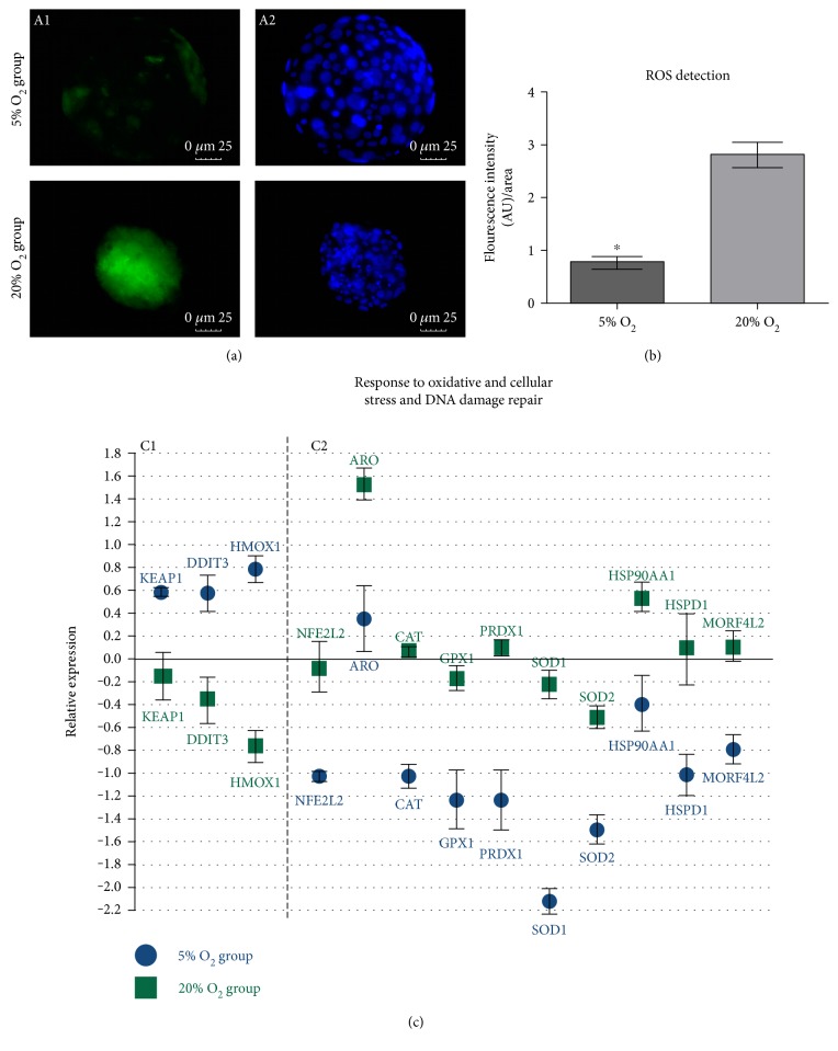
Figure 2.
(a) DCF probe detection in expanded blastocysts from the 5% O2 and 20% O2 groups (40x magnification); A1—fluorescence intensity of DCF in the presence of ROS; A2—Hoechst 33342 staining of the embryo cells nuclei. (b) Difference of fluorescence intensity generated by DCF probe between the analyzed groups. Mean ± SEM of the groups 5% O2 (0.76 ± 0.15) and 20% O2 (2.80 ± 0.18) (P = 0.001). (c) Genes related to oxidative and cellular stress response and DNA damage repair with difference between the analyzed groups; C1—genes upregulated in the 5% O2 group (P < 0.05); C2—genes downregulated in the 5% O2 (P < 0.05); and x-axis (0) represents the control sample.

