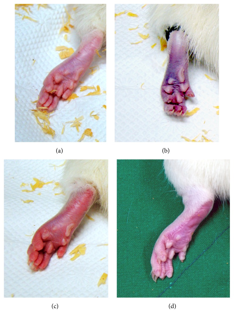
Figure 2.

Photographs of the left hind paw in chronic postischemic pain rats. (a) Normal hind paw. (b) Hind paw during ischemic event: a tight-fitting O-Ring has been placed just proximal to the ankle joint; the paw shows cyanosis. (c) Hind paw 10 minutes after reperfusion: the paw shows swelling and hyperemia. (d) Hind paw 3 days after reperfusion: swelling and hyperemia are decreased; the paw appears dry and shiny.
