
Figure 1.
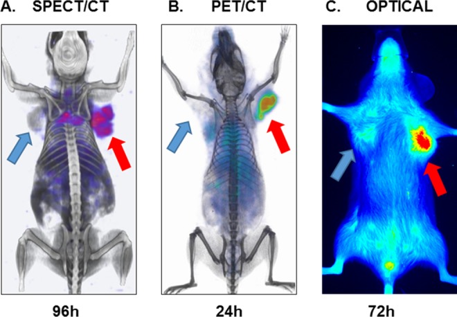
Imaging of tumor PD-L1 using atezolizumab: (A) SPECT/CT, (B) PET/CT, and (C) NIR imaging of PD-L1 using [111In]atezolizumab, [64Cu]atezolizumab, and NIR-atezolizumab, respectively, in NSG mice-bearing hPD-L1 tumor (red arrow) and control CHO (blue arrow) tumors. Adapted from Chatterjee et al10 and Lesniak et al11. CT indicates computed tomography; CHO, Chinese hamster ovary; PET, positron emission tomography; NIR, near-infrared; NSG, non-obese diabetic severe-combined immunodeficient gamma; SPECT, single-photon emission computed tomography.