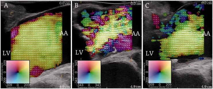
Figure 3.
Vector flow imaging of flow in the ascending aorta during the systole. (a) shows the systolic flow in a patient with normal aortic valve. (b) and (c) show systolic flow in a patient with aortic valve stenosis before (b) and after (c) valve replacement. Flow complexity is increased with aortic valve stenosis and reduced after valve replacement. LV = left ventricle. AA = ascending aorta. From Hansen et al.32 Reprinted with permission from Ultrasound in Medicine and Biology.

