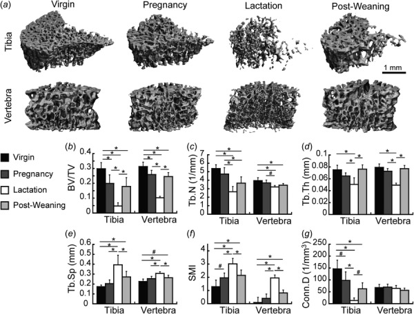
Fig. 1.

(a) Representative three-dimensional renderings of trabecular microstructure at the proximal tibia and the L4 vertebra in virgin, pregnancy, lactation, and 6-week post-weaning rats. (b)–(g) Microstructural parameters at the tibia and L4 at each reproductive stage, including: (b) BV/TV, (c) Tb.N, (d) Tb.Th, (e) Tb.Sp, (f) SMI, and (g) Conn.D. * indicate significant differences among groups (p < 0.05), # indicate trends toward differences among groups (p < 0.1).
