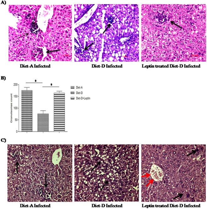
Figure 4.
Arrow indicates the structural organization and number of granulomas in H&E stained liver sections (20X). (A) Diet-A infected group with large and well-organized granulomas, Diet-D infected group with tiny granulomas, and leptin-treated diet-D infected group with well-organized granulomas. (B) Granulomas count; Diet-A, Diet-D and leptin-treated diet-D group. (C) Hepatic tissue degenerative changes; In the diet-A, infected groups showed, mild hepatic degeneration at the centrilobular region and proliferation of fibrous tissue at the peribiliary region. In the diet-D infected groups showed moderate to severe vacuolar degeneration was noticed at the periportal and centrilobular region. In the leptin-treated diet-D infected, most of the hepatocytes appeared normal, portal and periportal region along with the bile duct appeared normal, and mild hepatic degeneration was noticed in few places of the centrilobular region. Collective data of two independent experiments are shown.

