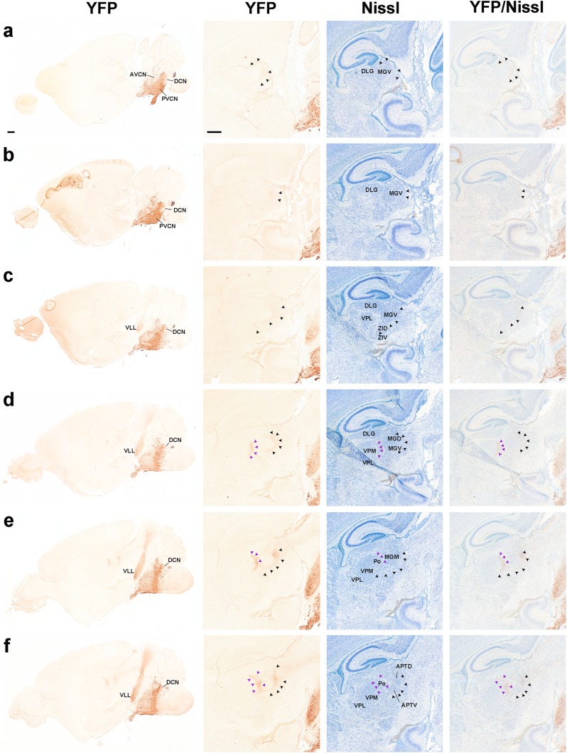
Fig. 13.
Illustration of r4-derived fibres projecting to the thalamus at P8. a–f Immunostaining with anti-GFP antibody on series of adjacent sagittal sections of a P8 b1r4-Cre/YFP brain, ordered from lateral to medial (left panels marked YFP in each set of four). High magnification of the thalamic region is shown in the next three images at each level, from adjacent sections stained with anti-GFP and Nissl and the corresponding merged YFP/Nissl images (right side images) to locate the r4-derived fibres projecting into the thalamus. The lateral lemniscal fibres (black arrowheads) course longitudinally roughly through the area occupied by the prospective medial, ventral and dorsal subdivisions of the medial geniculate nucleus in the thalamus. R4-derived trigeminothalamic fibres (violet arrowheads) project to the posterior thalamic nucleus and the medial part of the ventrobasal complex. For abbreviations see “list of abbreviations”. Scale bar 400 µm

