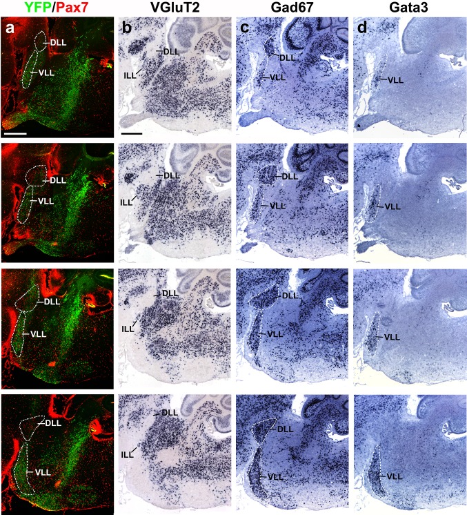
Fig. 16.
Series of adjacent lateral sagittal sections of a P8 b1r4-Cre/YFP brain stained with YFP/Pax7 (a), VGluT2 (b), Gad67 (c) and Gata3 (d). A dashed white line delimits the ventral (VLL), intermediate (ILL) and dorsal (DLL) nuclei of the lateral lemniscus. The DLL lies in r1 and is labelled by Pax7 and Gad67, being negative for VGluT2 and Gata3; this suggests a GABAergic/glycinergic phenotype of DLL. Separately, in the absence of Pax7 signal, YFP, Gad67 and Gata3 label the r4-derived VLL neurons in r2 and r3, which accordingly also have a GABAergic/glycinergic phenotype. Green fluorescent-labelled VLL neurons do not overlap with red-labelled DLL neurons, though green fibres pass through DLL. There is a vaguely delimited space between DLL and the massive VLL, where scarce YFP+/Gad67 +/Gata3 +/ neurons are found; in this zone, a group of VGluT2 + neurons form the ILL nucleus, which would accordingly consist of glutamatergic neurons. Scale bar 400 µm

