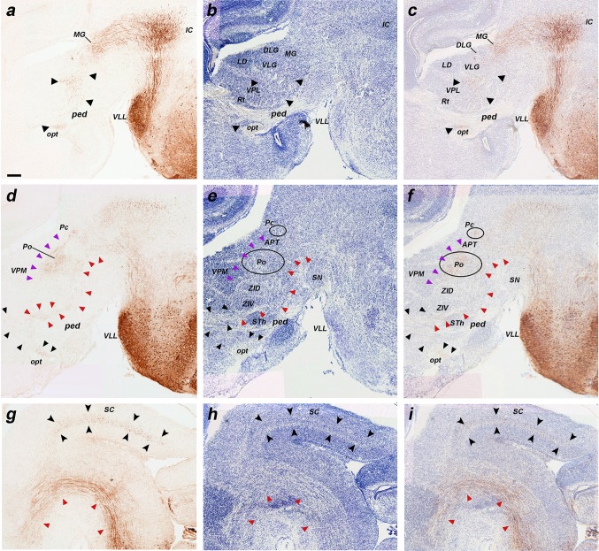
Fig. 8.
a–i Details at higher magnification of three of the sagittal sections of an E18.5 b1r4-Cre/YFP brain shown in Fig. 7, ordered from lateral to medial, and immunostained with anti-GFP (a, d, g), compared to Nissl-stained adjacent sections (b, e, h), and to merged YFP/Nissl images (c, f, i). The lateral lemniscus fibres penetrate profusely the inferior colliculus, entering radially its deep central part (a–c). A number of lemniscal fibres clearly extend through the brachium of the inferior colliculus (across midbrain and pretectum) to the thalamic medial geniculate nucleus (MG); more rostrally, the tract extends all the way to the supraoptic decussation (a–c, black arrowheads). These labelled lemniscal fibres course at the interface between the cerebral peduncle and the optic tract (opt, ped; rostralmost black arrowheads in a–c). Labelled fibres also penetrate the deep stratum of the superior colliculus (superficial to the periaqueductal grey) (SC; g–i, black arrowheads). The trigeminothalamic tract projects fibres to the posterior thalamic nucleus and the medial part of the ventrobasal complex (d–f, violet arrowheads). The medial tegmental tract crosses the tegmentum of the prepontine hindbrain, midbrain and diencephalon, reaching the hypothalamus (d–f, g–i; red arrowheads). For abbreviations see “list of abbreviations”. Scale bar 200 µm

