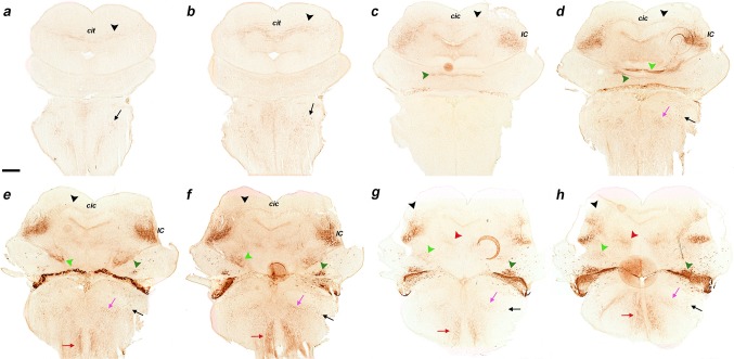
Fig. 9.
a–h Immunostaining with anti-GFP antibody on a series of horizontal sections through an E18.5 b1r4-Cre/YFP brain, ordered from dorsal to ventral. The YFP labels distinctly r4 and the r4 derivatives and related tracts. The lateral lemniscus (black arrowheads) reaches the deep part of the inferior colliculus (e, f) and extends fibres into the intercollicular commissure (c, d), the deep stratum of the superior colliculus (b, c) and the tectal commissure (a, b). The trigeminal cerebellopetal tract (light green arrowheads) reaches the cerebellum (e–h) and enters the cerebellar commissure (d). The vestibular cerebellopetal tract (dark green arrowheads) crosses the midline of the cerebellar nodule (c, d). Labelled fibres apparently derived from reticular and vestibular r4 neurons descend medially into the spinal cord forming the medial spinopetal tract (red arrows; e–h). The lateral vestibulospinal tract (black arrows) originates from r4-derived vestibular neurons (dark green arrowheads; e–h). The lateral trigeminal oro-spinal tract courses in an intermediate (lateral basal) tegmental position, intercalated between the medial spinopetal tract and the lateral vestibulospinal tract (pink arrows; d–h). For abbreviations see “list of abbreviations”. Scale bar 400 µm

