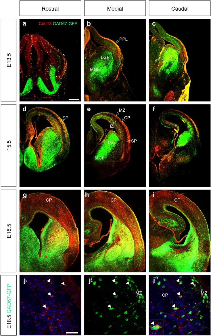
Fig. 3.
Expression of Cdh13 protein in cortical interneurons. Coronal sections from GAD67-GFP mouse forebrains at E13.5 (a–c), E15.5 (d–f) and E18.5 (g–i), were immunostained for Cdh13 (red) at rostral (a, d, g), medial (b, e, h) and caudal (c, f, i) levels. At E15.5 and E18.5, co-localization (yellow) is observed in the SP and CP. Hollow arrowheads indicate the positions of migratory streams of cortical interneurons. j, j″ Higher power images at E18.5; arrowheads indicate examples of interneurons that express Cdh13 protein (see inset). Scale bar in a 500 μm (a–i), in j 75 μm (j, j″). CP cortical plate, LGE lateral ganglionic eminence, MGE medial ganglionic eminence, MZ marginal zone, PPL preplate layer, SP subplate

