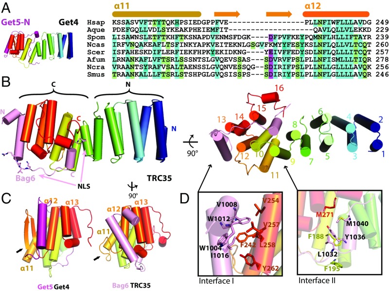
Fig. 1.
The structure of the Bag6NLS–TRC35 heterodimer. (A) The structure of Saccharomyces cerevisiae Get4–Get5N complex [Protein Data Bank (PDB) ID code: 3LKU], Get4 in rainbow and Get5 in magenta. Sequence alignment of TRC35/Get4 from Homo sapiens, Amphimedon queenslandica, Schizosaccharomyces pombe, Naumovozyma castelli, Saccharomyces cerevisiae, Aspergillus fumigatus, Neurospora crassa, and Sphaerulina musiva. The secondary structure based on fungal Get4s is highlighted above the text. (B, Left) The overall structure of Bag6–TRC35 heterodimer in cylinder representation with Bag6 in light pink and TRC35 in rainbow. The nuclear localization sequence is highlighted in sticks on Bag6. (B, Right) A 90° in plane rotated ‘bottom’ view. The 16 helices of TRC35 are labeled from N to C terminus. (C) Comparison of the C-terminal faces of TRC35 and Get4 that bind Bag6 and Get5, respectively. The arrows highlight the significant structural difference in the residues between α11 and α12. (D) Zoomed in view of the regions, defined as interface I and II. Hydrophobic residues that are involved in Bag6–TRC35 dimerization are shown as sticks.

