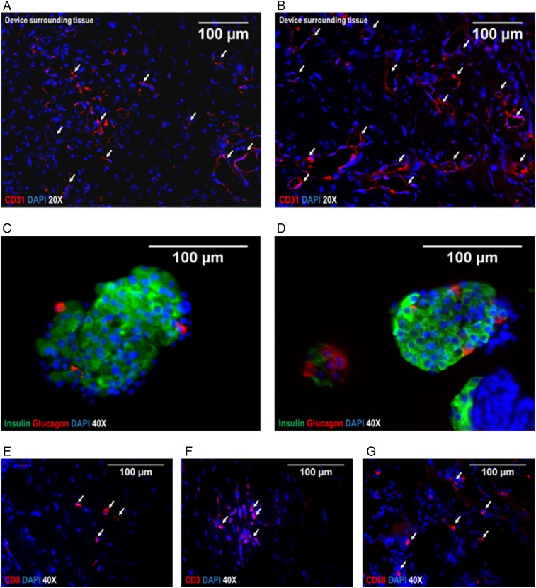
Fig. 4.
Immunohistochemical analysis of islets after explantation and device surrounding tissue. (A and B) Representative images of tissue surrounding the device on the peritoneal site, stained for CD31 to visualize strong vascularization as crucial for exchange of glucose/hormones. (Red) CD31, (blue) DAPI. (C and D) Representative images of porcine islets immobilized in alginate analyzed after explantation of the device at 6 mo. (Green) insulin, (red) Glucagon, (blue) DAPI. The architecture and cell composition of explanted islet grafts were not different to the structure at the time of implantation. Although beta cells are often found as single cells or grouped together to form the core of islets at younger age, the retired breeder animals as used in this study typically show a rather compact structure with beta cells scattered throughout the islet and alpha cells predominantly in the periphery. For analysis of local immune reactions at the transplantation site, the surrounding tissue was stained for CD8+ T-cells (E), CD3+ activated cytotoxic T-cells (F), and CD68+ macrophages (G); (red) respective CD-molecules, (blue) DAPI. Arrows indicate the relevant structures in the respective images (exemplary).

