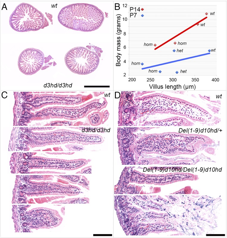
Fig. 5.
Reduced small intestine diameter and epithelial dysgenesis in Hoxd3hd and HoxDDel(1–9)d10hd milk-fed pups. (A) Two wild-type controls (Upper) and two Hoxd3hd-homozygous siblings (Lower) are shown. (B) The longest villi from at least six available sections from each individual were selected and photographed, and their longest axis was measured. In the diagram the body weight was plotted as a function of the average longest villus length. The Hoxd3hd stock at P14 is shown as red diamonds and a red line. The two diamonds on the right represent wild-type (wt) mice, and the two on the left represent homozygous (hom) siblings. A similar analysis was carried out with Del(1–9)d10hd at 7 d after birth and is depicted as blue diamonds and a blue line. The two values at the left represent homozygous animals (hom), the two in the middle represent heterozygous animals (het), and the one on the right is a wild-type control (wt). (C) H&E-stained paraffin sections of gut villi from wild-type control and two pairs of sections from two Hoxd3hd-homozygous specimens showing representative morphologies. (D) High-power photomicrographs of H&E sections of wild-type and heterozygous specimens and from two Del(1–9)d10hd-homozygous animals showing representative morphologies. (Scale bar, 1 mm in A and 100 µm in C and D.)

