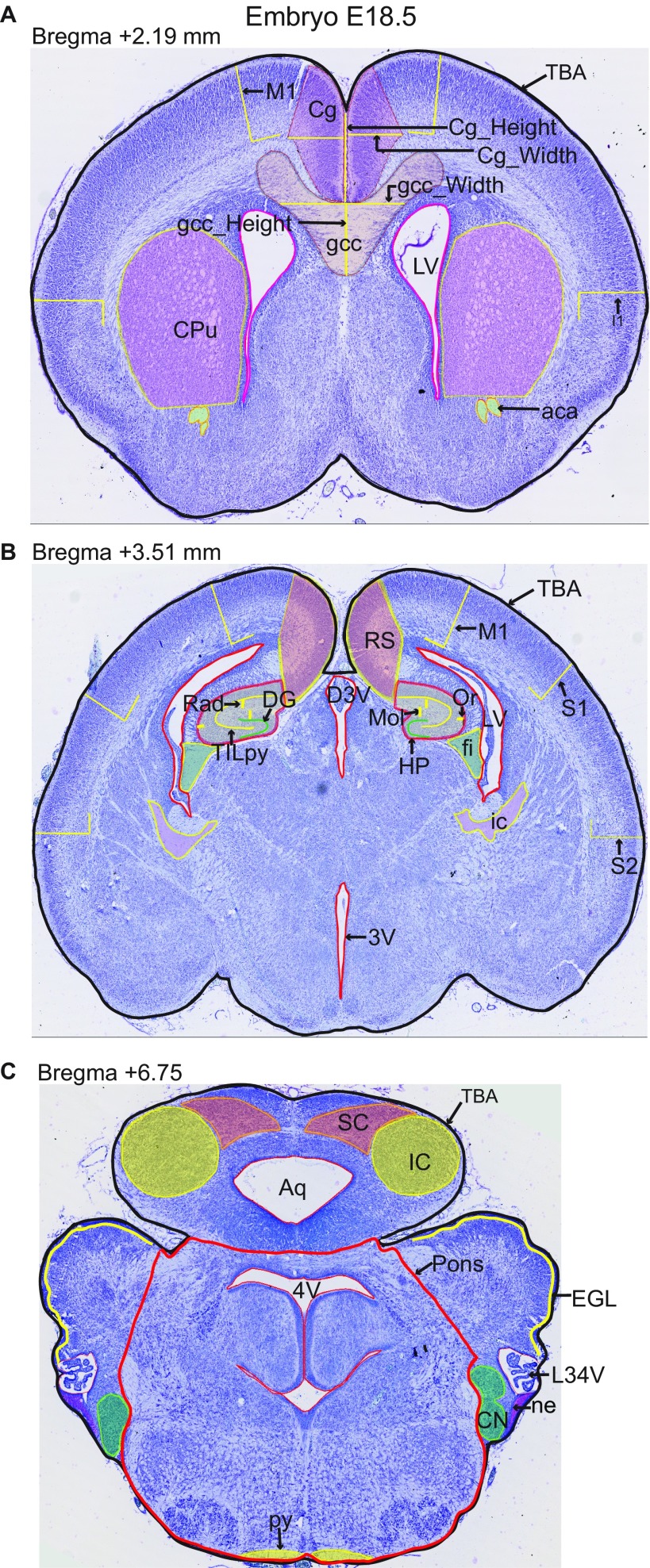
Fig. S5.
Coronal sections of interest at embryonic age E18.5. Regions quantified using ImageJ have been traced on representative images of the three coronal sections of interest. (A) Critical section 4 at 2.19 mm, (B) critical section 5 at 3.51 mm, and (C) critical section 6 at 6.75 mm. Parameter names are described in Dataset S8. All brain sections were stained using cresyl violet. 3V, 3rd ventricle; 4V, 4th ventricle; aca, anterior commissure; Aq, aqueduct; Cg, cingulate cortex; CN, cochlear nucleus; CPu, caudate putamen; D3V, dorsal 3rd ventricule; DG, dentate gyrus; EGL, external granule cell layer; fi, fimbria; Folia, number of folia; gcc, genu of corpus callosum; HP, hippocampus; I, insular cortex; IC, inferior colliculus; ic, internal capsule; LR4V, lateral recess of 4th ventricle; LV, lateral ventricules; M1, motor cortex; Mol, molecular Layer of HP; ne, neuroepithelium; Or, Oriens layer of HP; Pons, pons; py, pyramidal tract; Rad, radiatum layer of HP; RS, retrosplenial granular cortex; S1, primary somatosensory cortex; S2, secondary somatosensory cortex; SC, superior colliculus; T, top; TBA, total brain area; TILpy, total internal length of pyramidal layer. (Magnification: 20×.)

