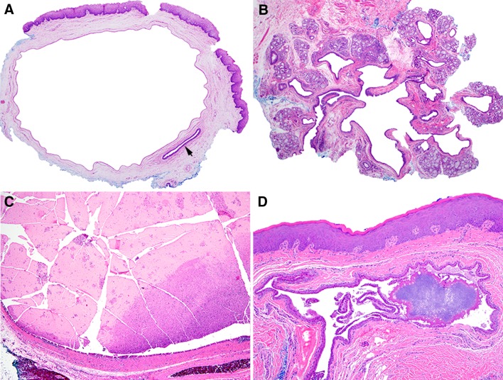
Fig. 2.
a Typical SDC with cystically dilated excretory duct, uninvolved excretory duct (arrow) is noted bottom right (×40). b SDC involving excretory duct as well as multiple smaller intralobular ducts located within distinct minor salivary gland lobules (×40). c SDC with intraluminal mucous plug (×40). d Early calcification occurring within longstanding SDC (×100)

