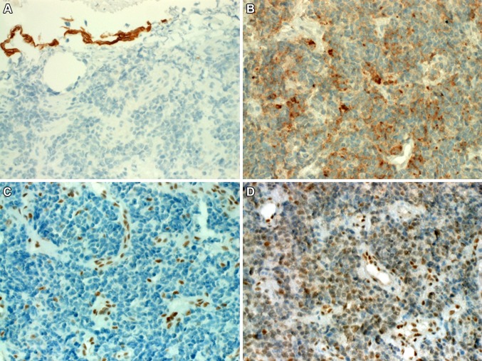
Fig. 2.
a The neoplastic cells were immunonegative with CK5 (surface epithelium was positive). b In this area of the tumor, weak to moderate punctate synaptophysin expression is seen. c Complete loss of SMARCA4 was seen in the neoplastic cells (normal endothelia as a control in the background). d SMARCA2 showed significantly reduced, albeit still recognizable nuclear staining in the tumor cells

