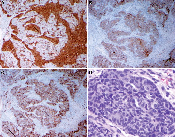
Fig. 4.
Two separate populations of cells are evident under immunohistochemistry. Low power: synaptophysin (a) strongly stains the more loosely cohesive cell population. These cells were also strongly positive for CD56. Pan-cytokeratin (b) and epithelial membrane antigen (c) distinctly highlight the solid component. Note when comparing the three images that separate cellular regions are highlighted, with the neuroendocrine population surrounding the epithelial marker-reactive cells. The epithelial-reactive solid component (d—high power) is composed of small cells with a high nuclear-to-cytoplasmic ratio and high mitotic activity

