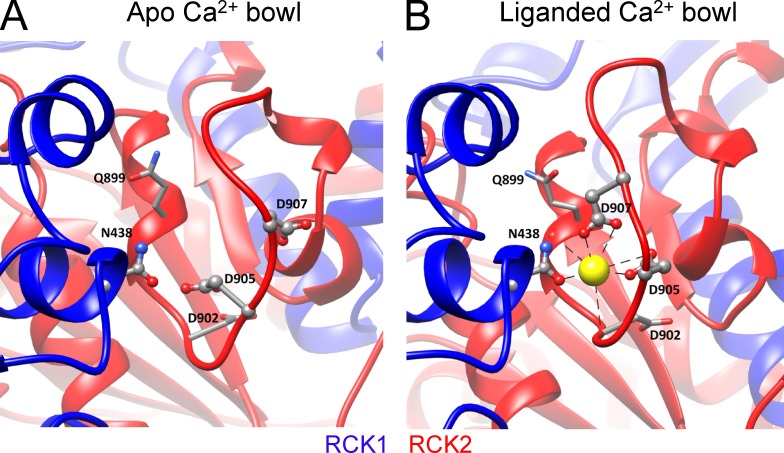
Figure 5.
The Ca2+ bowl. (A) High-resolution view of the metal-free aSlo1 structure from a portion of the RCK2 domain containing the Ca2+ bowl. Ca2+ coordinating residues are labeled (Aplysia numbering). Structures in A and B were aligned based on the αD helix in RCK1, which contains residue aN438 (mN449), proposed to provide a basis for cooperativity between Ca2+ binding in RCK1 and RCK2 of adjacent subunits. (B) View of the liganded aSlo1 structure showing Ca2+ coordination in the Ca2+ bowl. Residues that coordinate via backbone carbonyl are shown with stick side chains.

