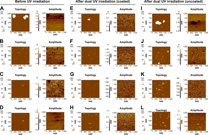
Figure 5.
EFM images of MO nanoparticles before/after dual UV irradiation.
Notes: MO nanoparticles before UV irradiation: (A) ZnO, (B) ZnTiO3, (C) MgO, and (D) CuO. MO nanoparticles after cyclic UV exposure using the coated area: (E) ZnO, (F) ZnTiO3, (G) MgO, and (H) CuO. MO nanoparticles after cyclic UV exposure using the uncoated area: (I) ZnO, (J) ZnTiO3, (K) MgO, and (L) CuO. MO nanoparticles were exposed to dual UV of UV-A and UV-C for 10 s over three cycles for 30 min. Topologies and amplitudes of MO nanoparticles were presented.
Abbreviations: EFM, electrostatic force microscopy; MO, metal oxide.

