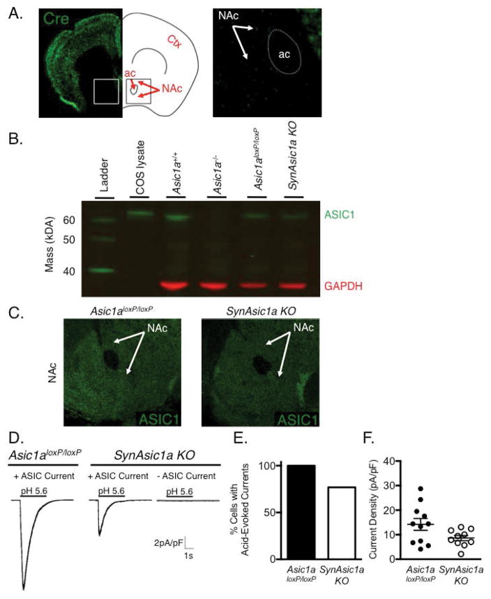
Figure 4. Preserved ASIC1A expression in the nucleus accumbens (NAc) of SynAsic1a KO mice.
A) Coronal section through the NAc of a SynRosa26 mouse. Although Cre-dependent eGFP expression was readily observed in cortical layers, it was rare in the NAc and striatum (Str). White square indicates magnified area including NAc (inset). Anatomic structures labeled for reference include cortex (Ctx) and anterior commissure (ac). B) Western blot of ASIC1A protein isolated from NAc punches of mice of the indicated genotypes, or COS cells transfected with Asic1a cDNA (Wemmie et al., 2002). ASIC1A (green, ~62 kDA) was detected in NAc from Asic1a+/+ and Asic1aloxP/loxP, and SynAsic1a KO mice with similar levels of ASIC1A expression in SynAsic1a KO mice and Cre negative Asic1aloxP/loxP controls. ASIC1A expression was absent in NAc of an Asic1a−/− mouse. GAPDH immunoblotting (red, ~37 kDa) was used to assess protein loading. C) ASIC1A immunohistochemistry of coronal sections through the NAc, showing similar levels of ASIC1A expression in SynAsic1a KO mice and Asic1aloxP/loxP controls. D–E) Whole-cell voltage-clamp recordings of NAc neurons in slice preparation. The majority of SynAsic1a KO NAc neurons exhibited acid-evoked currents and the proportion of neurons lacking acid-evoked currents did not differ significantly from Cre negative Asic1aloxP/loxP controls (n = 11, 13). F) Mean acid-evoked current density in SynAsic1a KO cells trended lower although it did not reach statistical significance.

