Figure 3.
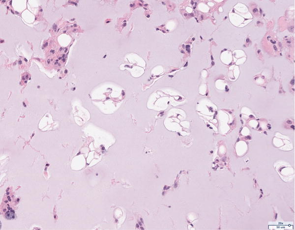
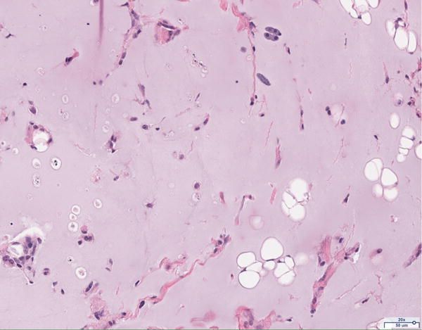
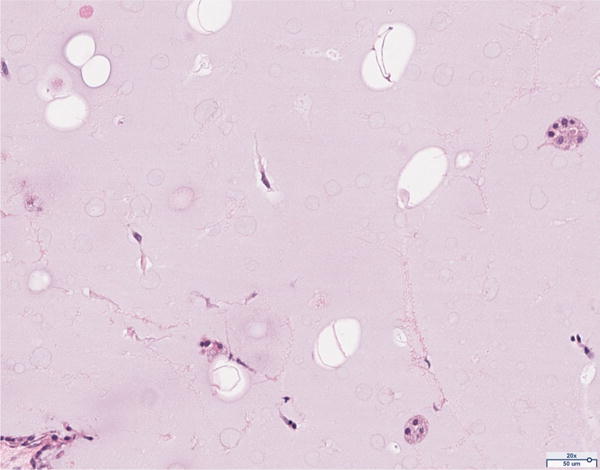
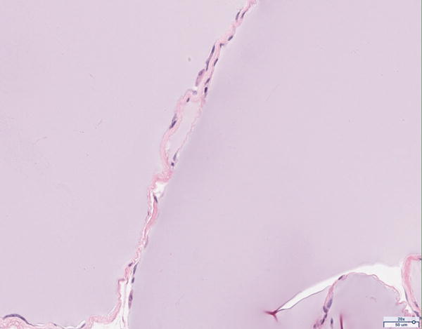
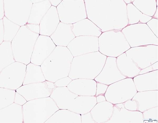
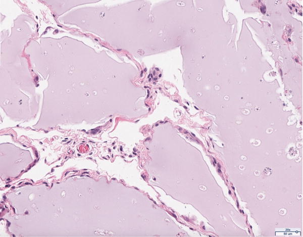
Hematoxylin and eosin (H&E) staining of explanted grafts. Grossly, the (a) 12 million cells per mL scaffold group demonstrated a greater degree of intra-scaffold adipocyte formation compared to the (b) 6 million cells per mL scaffold, (c) 1 million cells per mL scaffold, and (d) scaffold alone groups. Not surprisingly, the (e) fat alone samples displayed the greatest number of adipocytes. The 12 million group demonstrated increased formation of new ECM, fibrous tissue, and blood vessels (f).
