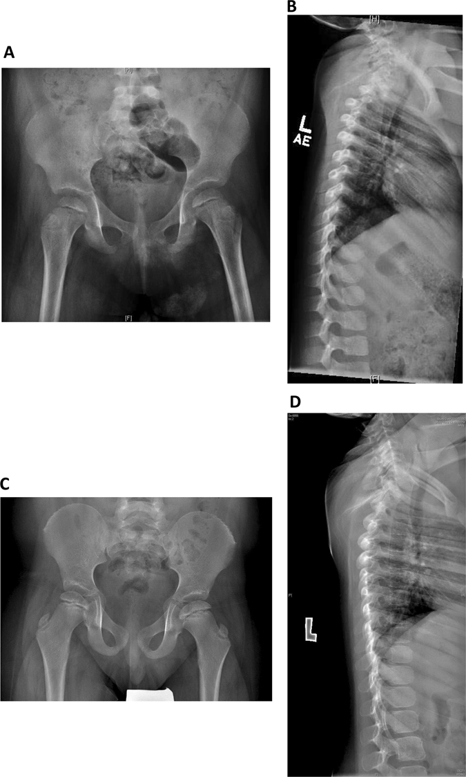
Fig. 2.

Skeletal features of Roifman syndrome in patients 1 and 2. a Pelvic X-ray of patient 1 featuring flattening of the humoral heads and shortening of the femoral necks, representing early stages of spondyloepiphyseal skeletal dysplasia. b Lateral spine X-ray of patient 1 featuring anterior vertebral notching of the lower thoracic vertebrae, and loss of lumbar lordosis. c Pelvic X-ray of patient 2 featuring bilateral small, flattened and slightly broadened femoral heads. d Lateral spine X-ray of patient 2 featuring loss of lumbar lordosis.
