Fig. 3.

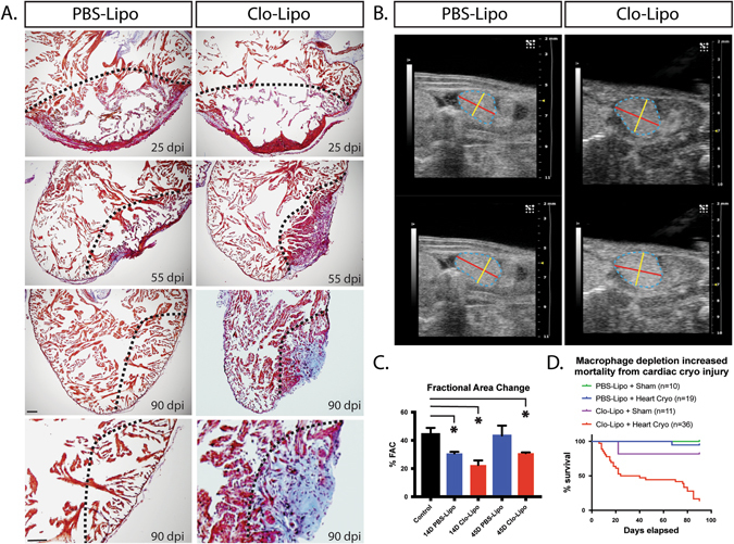
Macrophage depletion results in progressive scar formation and regenerative failure. a AFOG staining of Clo-Lipo macrophage depleted animals vs. PBS-Lipo control animals over the normal time-course of regeneration. Collagen deposition (blue) and fibrosis developed in the absence of macrophages and was not remodeled. b Ultrasound examination of the hearts post cryo-injury in macrophage depleted and normal animals. Representative end systolic (top) and end diastolic (bottom) images of the ventricles in the maximal longitudinal plane. Trace of the ventricle is shown in dashed blue line. Diameter marked with yellow and length with red overlays. c Assessment of cardiac performance based on calculations of % Fractional Area Change (%FAC). Cryoinjury results in a significant %FAC reduction in both Clo-Lipo and PBS-Lipo treated animals relative to uninjured control at 14 dpi. PBS-Lipo control animals regained normal function by 45 dpi while Clo-Lipo animal hearts remained compromised. d Clo-Lipo treatment altered animal survival post-cryo-surgery in two major phases. Scale bar = 500 μM
