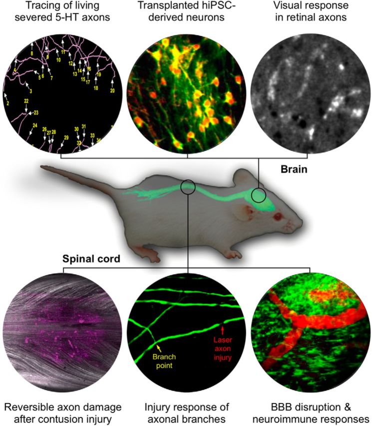
Figure 1.

In vivo imaging of CNS injury and disease. In vivo imaging of the brain and spinal cord has revolutionized our understanding of CNS pathology including axonal, vascular, and immune responses to CNS injury and disease. Topics illustrated here will be presented at a 2017 SfN Mini-Symposium. Top row, Tracing of living severed mouse serotonin axons in the neocortex (Jin et al., 2016), structural and functional in vivo imaging of transplanted human iPSC (hiPSC)-derived neurons expressing TdTomato (red) and GCaMP6 (green) (Real and De Paola, unpublished data), calcium imaging of visual response in retinal axons (Liang et al., unpublished data) are representative examples of imaging the dynamic responses of neuronal cells in the brain. Bottom row, Imaging the spinal cord after contusion injury (axons in white, cell death marker dye in magenta; Williams et al., 2014), injury response of axonal branches after laser axotomy (Lorenzana et al., 2015), and BBB disruption and microglia responses in autoimmune neuroinflammation (Davalos et al., 2012) illustrates how in vivo imaging allows the study of neuroimmune interactions and axonal responses to injury and disease. At center is an image of a mouse superimposed with the fluorescence image of a dissected central nervous system taken from a Thy1-XFP-transgenic mouse (modified from Misgeld and Kerschensteiner, 2006).
